|
| |
 |
| |
 |
| |
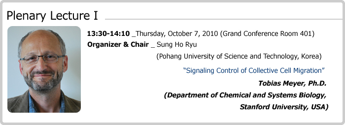
Professor Tobias Meyer is Professor of Chemical and Systems Biology in Stanford University since 1999. He received his MS in nuclear physics and then Ph.D in biophysics from the University of Basel, Switzerland. He trained as a Postdoctoral and Research Fellow at Stanford University with Professor L. Stryer. He had been a faculty member in Duke University Medical Center during 1991-1998. Professor Meyer has been working on the cell signaling, especially on the systemic understanding of signaling networks for cell migration and neuronal synapse formation and contributed over 100 publications in Cell, Science, Neuron etc. with pioneering research outputs. His current research direction may be summarized in the synopsis he described in his website as follows:
"Cells make use of an elaborate control system that integrates inputs from multiple receptors, computes this information and makes decisions about key cellular outputs such as cell migration, synapse formation, differentiation or proliferation. Our laboratory is focusing on discovering the rules that govern these decision processes by perturbing signaling steps, by monitoring signaling events and cell functions, and by employing mathematical modeling. We have already developed a number of novel biosensors and microscopy techniques to monitor cell signaling and functional processes over time, developed novel chemically induced enzyme activities for rapid signaling pathway perturbations and created a set of 2300 RNAi's to perturb signaling pathways by selectively reducing the expression of most human signaling proteins. We are particularly intrigued by the roles of calcium and lipid second messengers in the spatial and temporal coordination of cellular responses and a main current focus of the laboratory is on the questions how migrating cells polarize and chemotax and how neurons polarize their axons and dendrites and regulate their synapse number. While these studies focus on understanding specific control circuits, we are also working towards solving what is arguably the ultimate systems biology problem: How can a cells entire control system be quantitatively modeled? The experimental part of these studies makes use of genome-wide perturbations and monitoring multiple cellular decision points such as those triggering cell proliferation and differentiation. Our ultimate goals are to determine what elements of cellular control systems enable them to make specific decisions while being robust, how control systems have evolved, how we can synthetically create novel regulatory functions in existing control systems and how we can predict from mathematical models of signaling systems suitable molecular targets for therapeutic intervention”
|
 |
| |
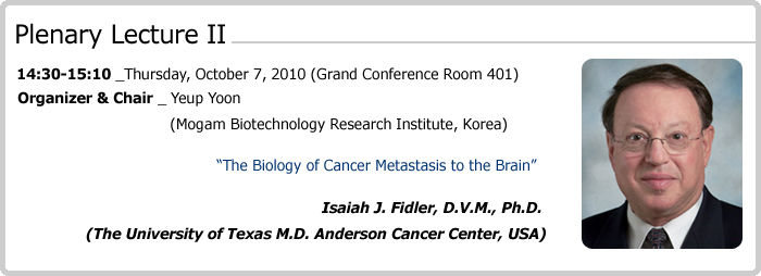
IIsaiah J. Fidler, D.V.M., Ph.D., is the R.E. ‘Bob’ Smith Distinguished Chair in Cell Biology, Professor and past Chairman Department of Cancer Biology, and Director of the Cancer Metastasis Research Center at The University of Texas M. D. Anderson Cancer Center, in Houston, Texas.
Over the past 40 years, Dr. Fidler has devoted his career to understanding the biology of cancer invasion and metastasis. In 1970, while carrying out research at the University of Pennsylvania School of Medicine, he described the first method for labeling tumor cells with the radioactive marker 125I-5-iodo-2'-deoxyuridine, allowing studies on the fate and distribution of tumor cells in vivo. These experiments provided the first evidence that only a small fraction of cells that enter the circulation survive to produce metastasis. Three years later, Dr. Fidler published a paper in Nature New Biology on the in vivo selection of successive tumor cell lines for metastasis, demonstrating that metastasis is a selective rather than random process-which up to then had been the prevailing view. In 1977, he published another landmark paper, this time in Science with his colleague Margaret Kripke at the NCI Frederick Cancer Research Center, demonstrating that clones derived in vitro from a parent culture of murine malignant melanoma cells vary greatly in their ability to produce metastatic colonies in the lungs of syngeneic mice. This work provided the first definitive evidence that tumors are biologically heterogeneous and that metastatic cells preexist in a cancer cell population and are not the result of adaptation during the metastatic process.
After moving from NCI-Frederick Cancer Research to The University of Texas, M.D. Anderson Cancer Center in Houston in the early eighties, he continued his work understanding the processes of tumor progression and biologic diversification of human malignancies. To this end, he has performed seminal work showing the utility of orthotopic nude mice models in studying the biology of human metastases compared with ectopic models, which generally fail to produce metastases. He has also demonstrated that specific organ microenvironments influence the resistance of metastatic cells to chemotherapy and experimental biological therapies.
His work on the importance of the organ microenvironment (e.g., in influencing tumor cell gene expression) has reinvigorated interest in the venerable ‘seed and soil’ hypothesis first put forward by the English surgeon Stephen Paget in 1889 (who noted the dependence of metastasis on cross talk between selected cancer cell ‘seeds’ and specific organ ‘soils’). More recently, Dr. Fidler’s group has shown that organ-specific cytokines (e.g., epidermal growth factor, vascular endothelial growth factor, transforming growth factor beta and platelet derived growth factor) also regulate angiogenesis in primary neoplasms and their metastases.
Dr. Fidler has authored more than 800 primary research articles, book chapters, and reviews. Among his many honors are the American Cancer Society’s Distinguished Service Award, City of Paris Medal of Vermeil, the WHO Medal for Biological Science, the Bristol-Myers Squibb Award for Distinguished Achievement in Cancer Research, and Nature Publishing’s Lifetime Achievement Award.
Much of what is known about the fundamental biology and mechanisms of metastasis originated from the Fidler group. Over 50 clinical fellows have passed through the doors of his laboratory, and he has been responsible for training and mentoring over fifty postdoctoral researchers, most of whom are leading research in the cancer field.
|
 |
| |
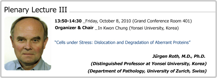
Professor Jűrgen Roth became the first Professor of Cell and Molecular Pathology in Europe when he was appointed to the University of Zurich in 1990. He directed the newly established Division of Cell and Molecular Pathology at the Department of Pathology until 2009 and currently is a Distinguished Professor at Yonsei University Graduate School, WCU program. He received his medical degree from the Friedrich Schiller University Jena and a PhD in Cell Biology from the University of Jena and of Basel in Switzerland. He trained as a Visiting Scientist at the Cancer Research Institute of the University of Vienna, Austria. He was a resident and docent in General and Surgical Pathology at University of Jena, Research Associate and Lecturer in the Department of Morphology at University of Geneva, and Associate Professor of Cell Biology at the Biocenter of the University of Basel. He has been Editor-in-Chief of the European Journal of Cell Biology and is currently Editor-in-Chief of Histochemistry and Cell Biology. He has served as president and past-president of the Society for Histochemistry and of the International Glycoconjugate Organization and as board member of the International Federation of the Societies for Histochemistry and Cytochemistry. He has been awarded a medical honorary degree from Peking University, was elected Honorary Member of the Society of Histochemistry, received medals of Honor from various Japanese Universities, was named Invited Fellow of the Japanese Society for Promotion of Science and was awarded many scientific prizes. Professor Roth has contributed over 300 publications to the cell biology of cellular organelles and for immunoelectron microscopy, has published or edited several books and organized many International Conferences and Symposia.
Professor Roth’s research interest encompasses the molecular cell biology of the endoplasmic reticulum and the Golgi apparatus in regard to protein glycosylation and protein quality control and of cell surface glycoconjugates during development and in tumors. He invented the immunogold technique for electron microscopy, the most powerful and most widely used technique of immunoelectron microscopy today and advised lectin-gold techniques as well. He was the first to directly demonstrate the subcompartmentalization of the Golgi apparatus and to characterize the TGN as part of the Golgi apparatus, to demonstrate that protein O-glycosylation starts in the Golgi apparatus, and for instance that polysialic acid of the neural cell adhesion molecule is an oncofetal antigen in human organogenesis and malignant tumors. His work on protein quality control resulted in the discovery of the first glyco-code for glycoprotein degradation by the ubiquitin-proteasome system, the identification of pre-Golgi intermediates as a major quality control check point, and led to the discovery of the first known ERAD dislocation receptor. His recent studies on the ERAD dislocation receptor EDEM1 established a novel vesicular exit pathway from the endoplasmic reticulum and demonstrated the importance of selective basal autophagy in quality control and protein turnover.
|
 |
| |
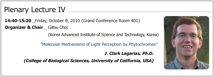
Professor J. Clark Lagarias received his Ph.D degree from University of California, Berkeley, joined the UC Davis faculty in 1980 as an assistant professor, and became the full professor in the section of Molecular and Cellular Biology in 1991. He was appointed as the Paul K. and Ruth R. Stumpf Professor of Plant Biochemistry from 1999 to 2006. Professor Lagarias was elected to the National Academy of Sciences (USA) in Plant Biology in May 2001.
Professor Lagarias has studied how plants perceive light through phytochromes, a group of plant photoreceptors. He is renowned for his research on the molecular and biochemical bases of how phytochromes perceive light information - light intensity and quality. His meticulous characterization of the biochemical and photochemical properties of purified phytochrome in 80’s presented a portrait of phytochrome as we know of it now. In late 80’s and early 90’s, he studied in planta synthesis of phytochrome chromophore and developed a method to assemble phytochrome holoproteins using recombinant apoproteins. The method provided scientists around the world a tool with which one can investigate the biochemical and molecular properties of phytochromes without grinding tons of etiolated oat seedlings in the cold dark room. In late 90’s, he showed that cyanobacteria has a phytochrome with histidine kinase activity and demonstrated that plant phytochromes possess serine/threonine kinase activity. These two discoveries startled the world, providing new insights into how phytochromes transmit light signal to their downstream signaling components. His revelations are going on in the 21st century as he recently engineered fluorescent phytochromes and light-independent constitutively active phytochromes. The achievements promise practical applications in biomedical and agricultural fields. The impression of his works reverberates in our minds, reminding us how much a brilliant and determined scientist can achieve in a mere three decades.
|
|
|
|
|

